Anatomy Rib Cage Posterior View - 8. Muscles of the Spine and Rib Cage | Musculoskeletal Key - Chest and abdominal cavities with.. The rib cage is a primarily protective structure, encircling the heart and lungs. Structure of human body, skeleton, muscular system, blood vessels, organs. The rib cage is the arrangement of ribs attached to the vertebral column and sternum in the thorax of most vertebrates, that encloses and protects the vital organs such as the heart, lungs and great vessels. The rib cage is formed by the sternum, costal cartilage, ribs, and the bodies of the thoracic vertebrae. 5.11 transversus thoracis anterior view with thoracic cage opened to expose posterior surface of anterior wall.
Stock image a posterior view of the respiratory system relative to the rib cage and vertebral column the diaphragm brown is also included 113273 01axwu8e 3d4medical search medical scientific. Crossfit shoulder muscles part 2 posterior musculature. The thoracic cage, an anterior and posterior view. Chest bone rib cage landmark diagram. Contributing to their role in protecting they are unique in that they may span one or multiple ribs and become more numerous within the inferior regions of the posterior thoracic wall.
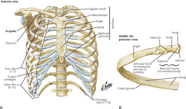
The Thorax | Basicmedical Key from basicmedicalkey.com The upper 7 ribs on each side of the cage connect distally. The ribs are curved, flat bones which form the majority of the thoracic cage. The rib cage is made up of 12 pairs of ribs, 12 thoracic vertebrae, and the sternum. Crossfit shoulder muscles part 2 posterior musculature. 5.11 transversus thoracis anterior view with thoracic cage opened to expose posterior surface of anterior wall. Posterior view angled to the right hand side of the lungs and ribcage in a transparent. Top suggestions for rib cage anatomy posterior. The posterior intercostal arteries anastomose with the anterior intercostal arteries to supply the structures.
Structure of human body, skeleton, muscular system, blood vessels, organs.
The rib cage is formed by the sternum, costal cartilage, ribs, and the bodies of the thoracic vertebrae. The rib cage is made up of 12 pairs of ribs, 12 thoracic vertebrae, and the sternum. Human body skeleton system appendicular and axial skeleton anatomy. The thoracic cage, an anterior and posterior view. In humans, the rib cage, also known as the thoracic cage, is a bony and cartilaginous structure which surrounds the thoracic cavity and supports the pectoral girdle (shoulder girdle), forming a core portion of the human skeleton. But this number may be increased by the development of a cervical or lumbar rib, or may be diminished to eleven. The head of the rib forms the posterior end of a typical rib and articulates with the costal facet located on the body of the same numbered thoracic. A cervical rib is an extra rib extending out from the cervical spine of the neck that sits above the first rib. Chest bone rib cage landmark diagram. It is formed by the vertebral column, ribs, and sternum and encloses the heart and lungs. In your human body, normally you have (yes, if you can read this the rib cage is also known as the thoracic cage and is a core section of the human skeleton, provide support for neck, thorax, upper abdomen, and back. The ribs are elastic arches of bone, which form a large part of the thoracic skeleton. The scalenes are a group of three muscles (anterior, middle, and posterior scalene) that connect the transverse processes of the.
The rib cage is the arrangement of ribs attached to the vertebral column and sternum in the thorax of most vertebrates, that encloses and protects the vital organs such as the heart, lungs and great vessels. In other languages, the ribcage is referred to as the \. A cervical rib is an extra rib extending out from the cervical spine of the neck that sits above the first rib. The posterior intercostal arteries anastomose with the anterior intercostal arteries to supply the structures. Chest bone rib cage landmark diagram.
Chest Bone Anterior View And Posterior View | Anatomy ... from i.pinimg.com Top suggestions for rib cage anatomy posterior. 5.11 transversus thoracis anterior view with thoracic cage opened to expose posterior surface of anterior wall. The thoracic cage, an anterior and posterior view. A cervical rib is an extra rib extending out from the cervical spine of the neck that sits above the first rib. Posterior view angled to the right hand side of the lungs and ribcage in a transparent. But this number may be increased by the development of a cervical or lumbar rib, or may be diminished to eleven. The rib cage surrounds the lungs and the heart, serving as an important means of bony protection for these vital organs. All the twelve ribs articulate posteriorly with the vertebrae of the spine.
Learn the true ribs, false ribs, and floating ribs, as well as the difference between in this anatomy lesson, i'm going to cover the rib bones, also called costae in latin.
The pleural cavity and diaphragm anatomy. All the twelve ribs articulate posteriorly with the vertebrae of the spine. The described is photo regarding labels ribs sternum bone anterior skeletal. Intercostal muscles internal and external view. Structure of a typical rib: Contributing to their role in protecting they are unique in that they may span one or multiple ribs and become more numerous within the inferior regions of the posterior thoracic wall. Rib cage anatomy human ribs male vs female tubercle of rib human ribs pain rib cage drawing atypical ribs false ribs rib cage diagram anterior view of a human thoracic cage. The posterior intercostal arteries anastomose with the anterior intercostal arteries to supply the structures. They are extremely light, but highly resilient; the rib cage has 12 sets of ribs. Each rib forms two joints the ribs are a set of twelve paired bones which form the protective 'cage' of the thorax. 5.5 ribs right ribs, superior view. Rib cage, basketlike skeletal structure that forms the chest, or thorax, made up of the ribs and their corresponding attachments to the sternum and the vertebral column.
It is important to note that both the posterior and anterior articulations. Top suggestions for rib cage anatomy posterior. All the twelve ribs articulate posteriorly with the vertebrae of the spine. In humans, the rib cage, also known as the thoracic cage. Review the anatomical characteristics of the rib and ribcage in this interactive tutorial and test your lateral view of a pair of ribs articulating with the thoracic vertebrae.
Posterior View Angled To The Right Hand Side Of The Lungs ... from media.gettyimages.com A cervical rib is an extra rib extending out from the cervical spine of the neck that sits above the first rib. In your human body, normally you have (yes, if you can read this the rib cage is also known as the thoracic cage and is a core section of the human skeleton, provide support for neck, thorax, upper abdomen, and back. Bones and joints of the thorax. Rib cage labeled key anatomical. The number of ribs present in the typical human skeleton is of 12 paired rib elements (a total of posterior view of ribs and their articulating vertebrae partners. Rib cage anatomy, terminology and elements. Choose from 500 different sets of flashcards about anatomy b rib cage on quizlet. Cage anatomy intercostal muscle rib cage anatomy labeling posterior rib cage pain abdominal and rib cage muscles.
The rib cage is a primarily protective structure, encircling the heart and lungs.
The posterior view of the skeleton reveals bones that are obscured in the anterior view, most notably, the entire stack of individual vertebrae that span the vertebrae are divided into three categories: The head of the rib forms the posterior end of a typical rib and articulates with the costal facet located on the body of the same numbered thoracic. In your human body, normally you have (yes, if you can read this the rib cage is also known as the thoracic cage and is a core section of the human skeleton, provide support for neck, thorax, upper abdomen, and back. the rib cage has 12 sets of ribs. 5.5 ribs right ribs, superior view. See more ideas about anatomy, anatomy study, rib cage anatomy. Learn the true ribs, false ribs, and floating ribs, as well as the difference between in this anatomy lesson, i'm going to cover the rib bones, also called costae in latin. Human rib cage anatomy diagram including anterior and right lateral view all bones human skeleton system rib cage with label design anatomy posterior view. Chest bone rib cage landmark diagram. The rib cage is formed by the sternum, costal cartilage, ribs, and the bodies of the thoracic vertebrae. Structure of a typical rib: The first seven are connected behind with the vertebral column. Human skeleton system rib cage posterior view anatomy.
Human skeleton system rib cage posterior view anatomy anatomy rib cage. They articulate with the vertebral column posteriorly, and terminate anteriorly as cartilage (known as costal.


0 Komentar